Onion Epidermal Cell Orientation . — strips isolated from the epidermis in the directions perpendicular and transverse to a net cellulose orientation can be used as an extensiometric model. — we found that the average orientation of cellulose microfibrils inside onion abaxial epidermal cell walls as. — atomic force microscopy (afm) can resolve cmf bundles and has been extensively utilized to image the innermost lamella in onion epidermal peel. peeled the onion wall ultrastructurally to get a close look inside an epidermal cell wall, visualizing its major wall polysaccharides. The epidermal cells of onions. In situ afm of cell walls undergoing extension demonstrated a variety of cmf movements, including reorientation, sliding, and kinking. These large cells from the epidermis of a red onion are naturally pigmented. — for more than 10 years epidermal cell layers from onion scales have been used as a model system to study the.
from www.alamy.com
— atomic force microscopy (afm) can resolve cmf bundles and has been extensively utilized to image the innermost lamella in onion epidermal peel. — for more than 10 years epidermal cell layers from onion scales have been used as a model system to study the. — we found that the average orientation of cellulose microfibrils inside onion abaxial epidermal cell walls as. — strips isolated from the epidermis in the directions perpendicular and transverse to a net cellulose orientation can be used as an extensiometric model. These large cells from the epidermis of a red onion are naturally pigmented. In situ afm of cell walls undergoing extension demonstrated a variety of cmf movements, including reorientation, sliding, and kinking. The epidermal cells of onions. peeled the onion wall ultrastructurally to get a close look inside an epidermal cell wall, visualizing its major wall polysaccharides.
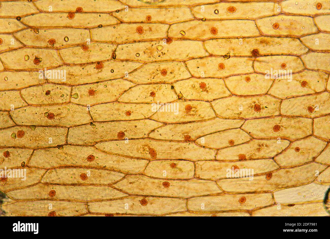
Epidermis of onion (Allium cepa) with cells, nucleus and walls
Onion Epidermal Cell Orientation In situ afm of cell walls undergoing extension demonstrated a variety of cmf movements, including reorientation, sliding, and kinking. — for more than 10 years epidermal cell layers from onion scales have been used as a model system to study the. These large cells from the epidermis of a red onion are naturally pigmented. peeled the onion wall ultrastructurally to get a close look inside an epidermal cell wall, visualizing its major wall polysaccharides. — atomic force microscopy (afm) can resolve cmf bundles and has been extensively utilized to image the innermost lamella in onion epidermal peel. In situ afm of cell walls undergoing extension demonstrated a variety of cmf movements, including reorientation, sliding, and kinking. — strips isolated from the epidermis in the directions perpendicular and transverse to a net cellulose orientation can be used as an extensiometric model. — we found that the average orientation of cellulose microfibrils inside onion abaxial epidermal cell walls as. The epidermal cells of onions.
From www.sciencephoto.com
Onion epidermal cells showing plasmolysis Stock Image B060/0059 Onion Epidermal Cell Orientation The epidermal cells of onions. In situ afm of cell walls undergoing extension demonstrated a variety of cmf movements, including reorientation, sliding, and kinking. — strips isolated from the epidermis in the directions perpendicular and transverse to a net cellulose orientation can be used as an extensiometric model. — we found that the average orientation of cellulose microfibrils. Onion Epidermal Cell Orientation.
From www.alamy.com
ONION SKIN CELLS (EPIDERMAL CELLS) / SHOWS CELL STRUCTURE AND NUCLEUS Onion Epidermal Cell Orientation — atomic force microscopy (afm) can resolve cmf bundles and has been extensively utilized to image the innermost lamella in onion epidermal peel. — for more than 10 years epidermal cell layers from onion scales have been used as a model system to study the. peeled the onion wall ultrastructurally to get a close look inside an. Onion Epidermal Cell Orientation.
From pixels.com
LM of cells in the epidermis of an onion Photograph by Science Photo Onion Epidermal Cell Orientation The epidermal cells of onions. These large cells from the epidermis of a red onion are naturally pigmented. — for more than 10 years epidermal cell layers from onion scales have been used as a model system to study the. In situ afm of cell walls undergoing extension demonstrated a variety of cmf movements, including reorientation, sliding, and kinking.. Onion Epidermal Cell Orientation.
From www.researchgate.net
Subcellular localization of AtTEM1 in onion epidermal cells. a, b Onion Onion Epidermal Cell Orientation peeled the onion wall ultrastructurally to get a close look inside an epidermal cell wall, visualizing its major wall polysaccharides. — strips isolated from the epidermis in the directions perpendicular and transverse to a net cellulose orientation can be used as an extensiometric model. — we found that the average orientation of cellulose microfibrils inside onion abaxial. Onion Epidermal Cell Orientation.
From cartoondealer.com
Micrograph Of Onion Epidermal Cells RoyaltyFree Stock Photo Onion Epidermal Cell Orientation — for more than 10 years epidermal cell layers from onion scales have been used as a model system to study the. — strips isolated from the epidermis in the directions perpendicular and transverse to a net cellulose orientation can be used as an extensiometric model. In situ afm of cell walls undergoing extension demonstrated a variety of. Onion Epidermal Cell Orientation.
From www.shutterstock.com
Onion Epidermal Cell Under Microscope Stock Photo 2210336617 Shutterstock Onion Epidermal Cell Orientation In situ afm of cell walls undergoing extension demonstrated a variety of cmf movements, including reorientation, sliding, and kinking. — atomic force microscopy (afm) can resolve cmf bundles and has been extensively utilized to image the innermost lamella in onion epidermal peel. These large cells from the epidermis of a red onion are naturally pigmented. — we found. Onion Epidermal Cell Orientation.
From www.sciencephoto.com
Epidermal cells of onion bulb Stock Image B060/0035 Science Photo Onion Epidermal Cell Orientation — we found that the average orientation of cellulose microfibrils inside onion abaxial epidermal cell walls as. The epidermal cells of onions. — atomic force microscopy (afm) can resolve cmf bundles and has been extensively utilized to image the innermost lamella in onion epidermal peel. In situ afm of cell walls undergoing extension demonstrated a variety of cmf. Onion Epidermal Cell Orientation.
From www.shutterstock.com
Onion Epidermal Cell Living Cell 400x Stock Photo 2221963957 Shutterstock Onion Epidermal Cell Orientation — atomic force microscopy (afm) can resolve cmf bundles and has been extensively utilized to image the innermost lamella in onion epidermal peel. These large cells from the epidermis of a red onion are naturally pigmented. In situ afm of cell walls undergoing extension demonstrated a variety of cmf movements, including reorientation, sliding, and kinking. — strips isolated. Onion Epidermal Cell Orientation.
From www.flickr.com
red onion epidermal cells, turgid photomicro Flickr Onion Epidermal Cell Orientation — atomic force microscopy (afm) can resolve cmf bundles and has been extensively utilized to image the innermost lamella in onion epidermal peel. In situ afm of cell walls undergoing extension demonstrated a variety of cmf movements, including reorientation, sliding, and kinking. These large cells from the epidermis of a red onion are naturally pigmented. peeled the onion. Onion Epidermal Cell Orientation.
From www.cell.com
Plant biology Peering deeply into the structure of the onion epidermal Onion Epidermal Cell Orientation — strips isolated from the epidermis in the directions perpendicular and transverse to a net cellulose orientation can be used as an extensiometric model. These large cells from the epidermis of a red onion are naturally pigmented. In situ afm of cell walls undergoing extension demonstrated a variety of cmf movements, including reorientation, sliding, and kinking. — atomic. Onion Epidermal Cell Orientation.
From cartoondealer.com
Micrograph Of Onion Epidermal Cells RoyaltyFree Stock Photo Onion Epidermal Cell Orientation The epidermal cells of onions. — strips isolated from the epidermis in the directions perpendicular and transverse to a net cellulose orientation can be used as an extensiometric model. — we found that the average orientation of cellulose microfibrils inside onion abaxial epidermal cell walls as. — for more than 10 years epidermal cell layers from onion. Onion Epidermal Cell Orientation.
From www.luc.edu
Onion Epidermis 100X General Biology Lab Loyola University Chicago Onion Epidermal Cell Orientation In situ afm of cell walls undergoing extension demonstrated a variety of cmf movements, including reorientation, sliding, and kinking. These large cells from the epidermis of a red onion are naturally pigmented. — for more than 10 years epidermal cell layers from onion scales have been used as a model system to study the. — atomic force microscopy. Onion Epidermal Cell Orientation.
From www.sciencephoto.com
LM of cells in the epidermis of an onion Stock Image B060/0029 Onion Epidermal Cell Orientation — atomic force microscopy (afm) can resolve cmf bundles and has been extensively utilized to image the innermost lamella in onion epidermal peel. In situ afm of cell walls undergoing extension demonstrated a variety of cmf movements, including reorientation, sliding, and kinking. peeled the onion wall ultrastructurally to get a close look inside an epidermal cell wall, visualizing. Onion Epidermal Cell Orientation.
From www.researchgate.net
Localization of GhSOS1 in onion epidermal cells. AC Onion epidermal Onion Epidermal Cell Orientation The epidermal cells of onions. In situ afm of cell walls undergoing extension demonstrated a variety of cmf movements, including reorientation, sliding, and kinking. — atomic force microscopy (afm) can resolve cmf bundles and has been extensively utilized to image the innermost lamella in onion epidermal peel. — strips isolated from the epidermis in the directions perpendicular and. Onion Epidermal Cell Orientation.
From schematiclistboons88.z13.web.core.windows.net
Onion Cell Diagram Labeled Onion Epidermal Cell Orientation peeled the onion wall ultrastructurally to get a close look inside an epidermal cell wall, visualizing its major wall polysaccharides. — strips isolated from the epidermis in the directions perpendicular and transverse to a net cellulose orientation can be used as an extensiometric model. These large cells from the epidermis of a red onion are naturally pigmented. The. Onion Epidermal Cell Orientation.
From www.alamy.com
High resolution light photomicrograph of Onion epidermus cells seen Onion Epidermal Cell Orientation — we found that the average orientation of cellulose microfibrils inside onion abaxial epidermal cell walls as. — atomic force microscopy (afm) can resolve cmf bundles and has been extensively utilized to image the innermost lamella in onion epidermal peel. — strips isolated from the epidermis in the directions perpendicular and transverse to a net cellulose orientation. Onion Epidermal Cell Orientation.
From www.pinterest.co.kr
Epidermal onion cells under a microscope. Plant cells appear polygonal Onion Epidermal Cell Orientation — for more than 10 years epidermal cell layers from onion scales have been used as a model system to study the. — atomic force microscopy (afm) can resolve cmf bundles and has been extensively utilized to image the innermost lamella in onion epidermal peel. peeled the onion wall ultrastructurally to get a close look inside an. Onion Epidermal Cell Orientation.
From www.alamy.com
ONION SKIN CELLS (EPIDERMAL CELLS) SHOWS CELL STRUCTURE AND NUCLEUS Onion Epidermal Cell Orientation — strips isolated from the epidermis in the directions perpendicular and transverse to a net cellulose orientation can be used as an extensiometric model. The epidermal cells of onions. — atomic force microscopy (afm) can resolve cmf bundles and has been extensively utilized to image the innermost lamella in onion epidermal peel. In situ afm of cell walls. Onion Epidermal Cell Orientation.
